top of page
DRUG DISCOVERY / CELL BIOLOGY
Drug discovery is a crucial element in the pharmaceutical industry, and biosensors are just now being utilized to uncover perfect drugs to the deadly diseases. In addition, experiments using biosensors are showing how they can detect analytes and shed light on cell systems and interactions. Drug discovery is the process of analyzing multiple molecules that could possibly cure certain diseases while cell biology is the study of life in cells. Please continue reading to learn about how biosensors are being employed to discover future drugs!
In vivo imaging is a technique used in live animals to view certain molecules that are inside of the body. Using quantum dots, or fluorescent nanoparticles, researchers are able to track down disease agents and signals, and use that information for drug development. When a certain molecule or reaction is present, the fluorescent particle is activated and glows in correlation to the quantity of whatever is being measured. Thus, in vivo imaging serves as an early detection system for diseases, as well as monitoring the disease stages in the host. Fluorescent biosensors are most commonly paired with in vivo imaging to locate the specific molecules due to their precision and speed. One disadvantage of using these biosensors is that while the temporal resolution, or the amount of precision in a short amount of time is good, the spatial resolution, or the image quality is not. However, researchers are working currently to produce higher quality photos by improving the existing biosensors. Below are some examples of biosensors in in vivo imaging:
BIOSENSORS IN IN VIVO IMAGING

The green fluorescent protein (GFP) coupled with a biosensor emits light when it detects activation of certain proteins. GFP was first discovered in the jellyfish species Aequorea victoria; whenever another protein called aequorin would detect Ca+ ions and show a flash of blue, GFP would be activated into producing green light.

The FRET-based biosensor can use fluorescence to monitor interactions between proteins and DNA. They can also detect structural protein changes as well. FRET stands for Forster (or fluorescence) Resonance Energy Transfer, which is the process of energy transfer between two fluorophores, or fluorescent chemical compounds.

Naturally found in fireflies, bioluminescent luciferase is an oxidative enzyme used to detect studying cell populations and their particular functions, such as cellular ATP levels. Luciferase biosensors can also sense the apoptosis, or self-destruction of cells, triggered by Mycobacterium tuberculosis proteins.
BIOSENSORS IN DRUG DISCOVERY
Drug discovery, as the name suggests, deals with finding certain molecules that can be used to resist and fight particular diseases. Drug candidates can be identified, characterized, optimized, and screened by using several methods. The most common of these methods are assays, which are procedures that detect specific analytes in cells. Another method utilized in drug discovery is the High Throughput Screening (HTS) which has a high affinity for specific compounds by employing labels. Unfortunately, labels, or reporter elements which are fluorescent, luminscent, radiometric, or calorimetric in obtaining measurements, can interfere with molecular interactions and produce false data. In order to prevent changes in the cell biology of target receptors, biosensors have been added in the mix of tools for drug discovery.
Biosensors can help drug developers study systems cell pharmacology. They can detect toxicity levels, which is important since there is always a potential risk that a drug candidate has unplanned dangerous side effects.
Biosensors can make the drug discovery less difficult and more efficient as well, since biosensors require low maintenance and less processed samples. Contrary to having multiple assays for each possible outcome of the signaling pathway, now only one assay and biosensor combo can study all the effects.
The biological and transducer component in a biosensor make an ideal configuration for the study between a drug and the immobilized biological compound. Biosensors coupled with cell-based assays can not only sense drug potentials, but can also activate pathways for toxicity tests. Biosensors are useful for probing cell signalling as well, for they can record the responses living cells send when various drugs or toxins approach and infiltrate them.
Previously used in biomolecular interaction analysis, label-free, optical biosensors have recently been applied in biochemical and cell-based assays rather than the fluorescent biosensors mentioned above in in vivo imaging. This is because label-free biosensors do not use sensitive dyes as reporters of biomolecular interaction, which can harm the cells' chemical composition.
While labeled technologies only verified the presence of the target molecule, label-free biosensors can obtain valuable information of binding affinity as well as how an analyte binds during interaction by perceiving subtle mass and heat capacity changes from the surface of the sensor substrate. Thus, label-free biosensors actually enhance the entire drug discovery process! Learn about a few detailed examples below!


BIOSENSORS IN CELL BIOLOGY
Biosensors are also employed in discovering new pathways in cell biology, which is a field that studies the anatomy and processes of a cell. How analytes bind and the affinity for it can reveal new information on existing, vague processes. Like drug discovery, cell biology typically uses optical biosensors to measure cell responses. This is because cell biology examines the same parameters as drug discovery, but encompasses target molecules not applicable as drug candidates. These optical biosensors use either surface plasmon resonance (SPR) or resonant waveguide grafting (RWG) to help monitor pathways with high temporal resolution, which is the preciseness of a measurement with respect of time.
To learn more about SPR or RWG, click .

Biosensors can help detect cell adhesion in the extracellular matrix (ECM) when cells naturally bind to the surface of the biosensor. Learning about cell adhesion is important because cells communicate through cell adhesion. Cells base their behavior on cell binding, which makes adhesion is vital for survival and proper function of the cell.

Scientists can gain more insight into cell-to-cell communication through biosensors. Cells connected by gap junctions can pass messengers such as cyclic AMP through the cytosol to communicate with their neighbors. Biosensors can trace specific toxin and killers that harm the cells by checking whether the communication is being hindered or not.

Biosensors can also detect cell signaling, a crucial mechanism of the cell to recieve data from outside and create a response. Biosensors can detect G-protein coupled receptors (GCPRs), and growth -factor receptor tyrosine kinases (RTKs). Their discovery can prompt drugs to be made against them, for if the growth receptor is destoryed, the malignant cell will die.
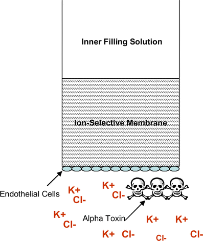
Non-invasive biosensors can characterize cell barrier functions in the cell. Two cell barriers that have been tested for permeability have been the blood brain barrier (BBB) and the epithelial cell barrier (ECB). By having biosensors measure permeability, reseachers can figure out the function of these barriers and see how selective they are. Learn more

The BBB protects the central nervous system due to its highly selective permeability. The BBB also stabilizes the brain for important neuron signals. The ECB regulates homoeostatis through its tight juctions between cells.
Electric biosensors are most commonly used due their sensitivity to charged ions.
(W-66)
(W-67)
(W-68)
Here's a summary of the cell interactions that biosensors can detect:
(W-69) to (W-71)
(I-90) to (I-92)
(I-93) to (I-95)
(I-96) to (I-98)
(I-99) to (I-103)

(I-104)
bottom of page